Gram Staining
Goal
Assess whether the biological sample contains Gram positive or Gram negative strains.
Materials
- Gas burner
- Crystal Violet
- 95% alcohol
- Iodine
- Safranin
- Tap water
- Blotting paper
Method
- Prepare a drop of sample on your microscope slide according to our Method
- Leave the sample to air dry for 5 to 10 minutes.
- Fixate by moving the slide back and forth through a flame for a few seconds.
- Stain the sample by a droplet of crystal violet and let it stain for max 60 seconds.
- Wash the crystal violet by tap water.
- Add the iodine solution for 60 seconds.
- Wash with tap water.
- Decolorize using 95 % ethanol and immediately rinse with tap water. Don’t let it on the slide for too long.
- Counter stain with safranin for about 60 seconds.
- Wash of the stain with tap water.
- Remove excess water using the blotting paper.
- Cover the sample with a cover slide.
- Take a look with the microscope with full light intensity (open diaphragma)
Interpreting the results
- Gram positive cells appear violet
- Gram negative cells appear red
Composition of the stains
- Crystal violet: 1 g of dry crystal violet in 10 mL 95% ethanol
- Iodine solution: 1 g of iodine in 100 mL water
- Safranin: 0.25 g of safrin in 10 mL 95% ethanol and 90 mL water
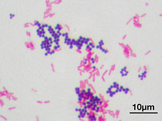
Microscopic image of a Gram stain of mixed Gram-positive cocci (Staphylococcus aureus ATCC 25923, purple) and Gram-negative bacilli (Escherichia coli ATCC 11775, red). Magnification: 1.000x - Picture by Y tambe CC BY SA 3.0 license
Read more
Back to BHA2 - Class 4