Loeffler Staining
Goal
Visualize cell nuclei in eukaryote cells.
Materials
- Gas burner
- 95% alcohol
- Loeffler’s methylene blue solution
- Tap water
- Bucket or beaker glass
- Blotting paper (alternatively: Tork roll or toilet paper)
- Clear nail polish
- Acetone
- Cotton swab
Composition of the stain
- Methylene Blue 3.0 g
- Potassium Hydroxide KOH, 10% 1.0 ml
- Ethanol, 95% 300.0 ml
Method
- Prepare a drop of sample on your microscope slide according to our Method
- Air dry the sample for 5 to 10 minutes.
- Soak the cells in a drop of 95% alcohol to dehydrate the cells
- Air dry the sample for 5 to 10 minutes, make sure all the alcohol is evaporated
- Fixate the cells by quickly moving the object glass through a flame.
- Let the sample cool down.
- Add the Loeffler’s solution for about 60 seconds.
- Remove excess stain by bathing / dipping your glass slide into a beaker glass / bucket with tap water.
- Remove excess water using blotting paper.
- Let the slide air dry to get rid of the remaining water.
- Put 2 drops of clear nail polish onto the sample.
- Cover the sample with a cover slide.
- Try to get rid of air bubbles by putting light pressure on the cover slide.
- Remove excess nail polish with a cotton swab with acetone.
- Take a look at the sample through the microscope with the diaphragm fully open.
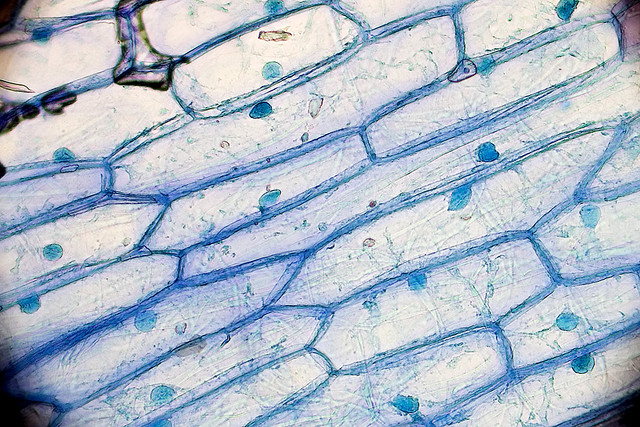
Microscopic image of onion cells stained with methylene blue, so the nuclei are visible. Magnification: 100x. - Picture by Umberto Salvagnin CC BY 2.0 license
Read more
Back to BHA5 - Class 4